RIAM uses the latest in imaging technology including 3 Tesla Magnetic Resonance Imaging (3T MRI), Breast MRI, Multi Detector Computed Tomography (MDCT), Intravascular Ultrasound (IVUS), Digital Mammography and CAD and Digital Radiography (DR). Below you can read about the different technologies and learn more about the procedures we perform. You can also find more information about diagnostic and interventional radiology at the following websites:
DIAGNOSTIC RADIOLOGY
Digital Radiography and Fluoroscopy
Digital Radiography uses a small amount of radiation to produce images of body tissue without film. Radiographs (x-rays) are useful in examining the lungs, heart, an air filled bowel and bones. Fluoroscopy is a type of real-time x-ray imaging used to provide movement. Radiologists can view specific body parts while they are functioning, and this is a common way to examine swallowing functions, the stomach, and the small and large bowel.
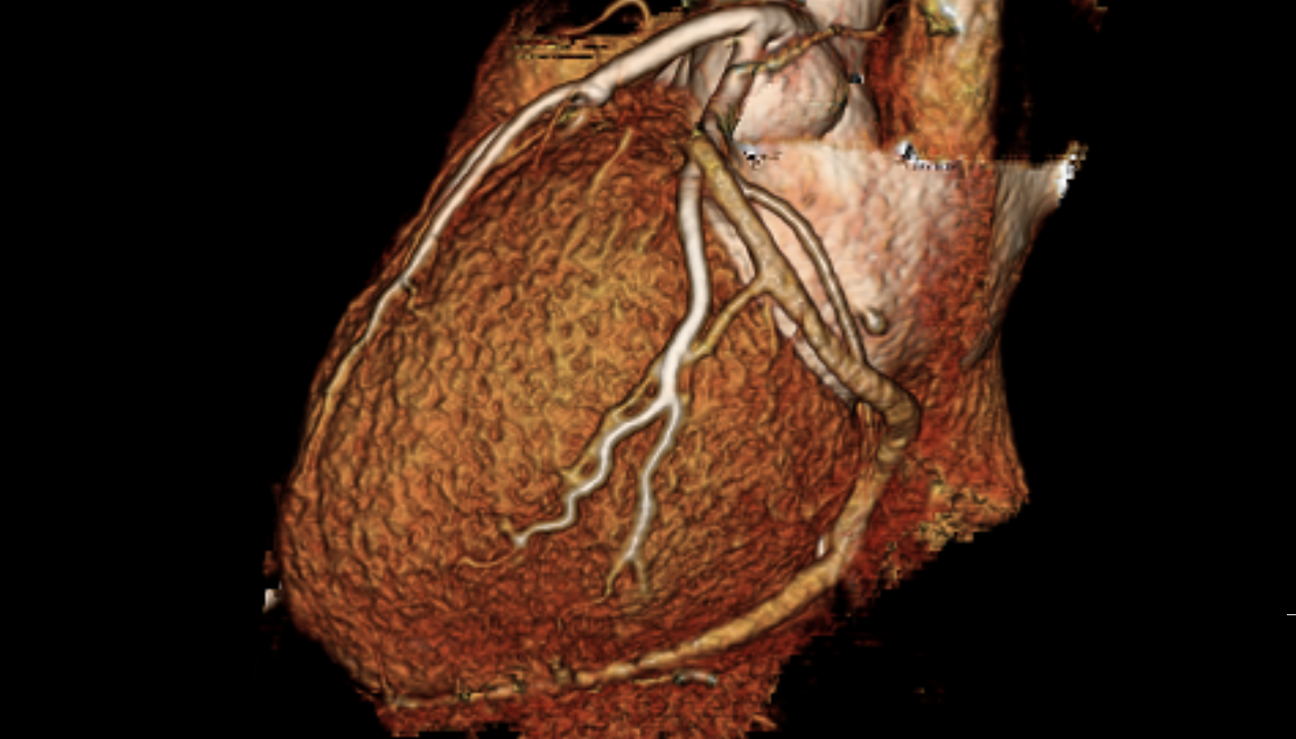
CT, CAT (Computed Tomography, Computed Axial Tomography)
CT scanning uses specialized x-ray generating and detecting equipment with a computer to produce multiple images of the inside of a body. The computer generates an axial image that can be as thin as 0.6 or as thick as 5 mm.
Ultrasound
Ultrasound uses high-frequency sound waves and their reflections to produce real time images of internal organs, movement within the organs, and blood flow through vessels. Ultrasound exams do not use ionizing radiation.
MRI (Magnetic Resonance Imaging)
Using a super strong magnetic field, radio waves and a computer, the MRI produces detailed cross-sectional images of the body. The MRI scan can obtain images in any predetermined plane (axial, sagittal, coronal, or oblique). MRI does not use ionizing radiation.
Nuclear Medicine
Nuclear Medicine utilizes a low risk amount of radioactive substance called a radionuclide, which is injected intravenously, intrathecally, or inhaled to view the physiological function of an organ or system. This type of imaging is useful for detecting tumors, infections, internal bleeding, blood cell disorders and functioning or non-functioning of organs.
Digital Mammography with CAD (including 3D/tomo)
Digital mammography and CAD (Computer-Aided Detection), are utilized to perform and interpret x-ray examination of the breasts. Mammography can detect abnormalities too small to be felt.
Minimally Invasive Cancer Treatments
Interventional radiologists are board-certified physicians who deliver minimally invasive treatments with less risk, less pain and less recovery time than traditional surgery to treat cancers. The minimally invasive treatments listed below are treatment options for people who are not candidates for surgical treatments, those who don’t want surgery and individuals whose cancer did not respond to cancer drugs.
- Chemoembolization involves the delivery of cancer-killing medication through a catheter (a thin, flexible tube) directly to the affected organ, decreasing blood flow in the arteries that supply the cancer.
- Selective Internal Radiation Therapy (SIRT), also known as Y-90, treats cancer through the injection of small spheres of radioactive substance into the blood vessels supplying the tumor.
- Microwave ablation is a thermal ablation technique that uses electromagnetic waves to destroy cancer cells.
- Palliative interventional radiology provides cancer patients with pain relief and symptom management when the patients enter into the terminal phase of their illness.
Uterine Fibroid Embolization
UFE involves inserting microspheres into the blood vessels of the fibroids, reducing blood flow and causing them to shrink. It is an alternative to a hysterectomy for patients with chronic pelvic pain and heavy bleeding. To contact our group directly about UFE, click here.
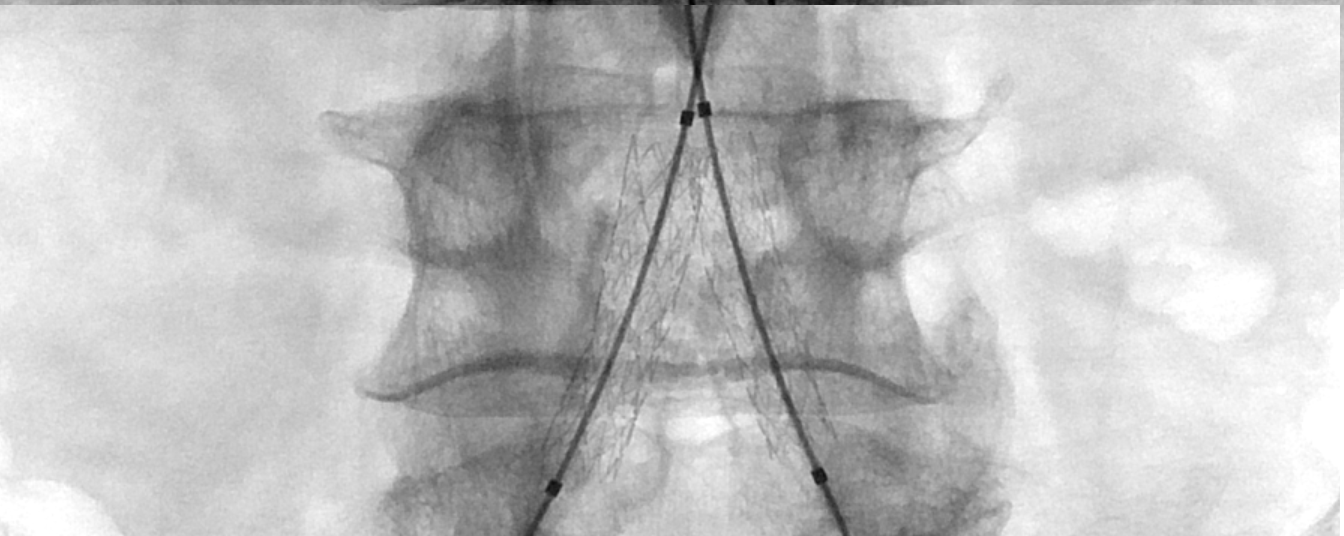
Vertebroplasty/Kyphoplasty
Vertebral body augmentation helps treat pain and stabilize vertebral compression fractures. Typically done on an outpatient basis, it involves making a tiny incision in the skin while using image guidance to insert a needle filled with specially formulated acrylic bone cement into the vertebra.
Deep Vein Thrombosis (DVT) Thrombolysis and Stenting for Acute and Chronic DVT
Catheter-directed thrombolysis is performed under fluoroscopic guidance. This procedure is designed to break up the clot, restore blood flow within the vein, and preserve valve function. Narrowing in the vein that might have caused clot formation can be treated by an interventional radiologist with a balloon venoplasty or stent placement.
Inferior Vena Cava (IVC) Filter Placement and Removal of Temporary IVC Filters
For patients with DVT of the legs who cannot tolerate thrombolysis or medical therapy, an IVC filter may be inserted. IVC filter placed within the IVC that traps blood clots preventing them from traveling to the lungs and causing a pulmonary embolism (PE), but allows normal liquid blood to pass.
Vascular Access including Subcutaneous Ports and Dialysis Catheters
Venous access is obtained through a small tube that is inserted beneath the skin providing a simple, pain-free way to perform dialysis or draw blood or give medication or nutrients. This spares the patient the irritation and discomfort of repeated needle sticks.
You can find additional minimally invasive treatments we perform here.